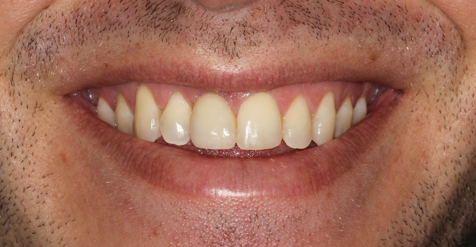
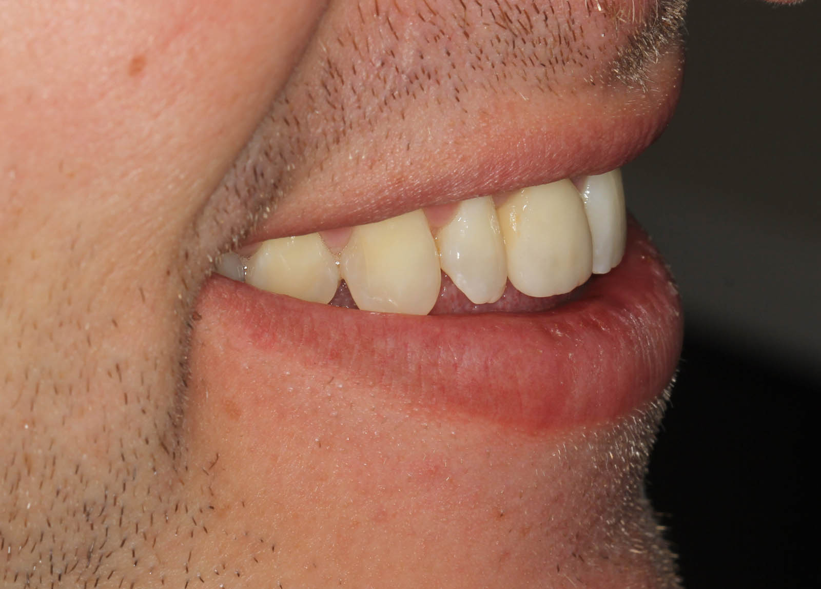
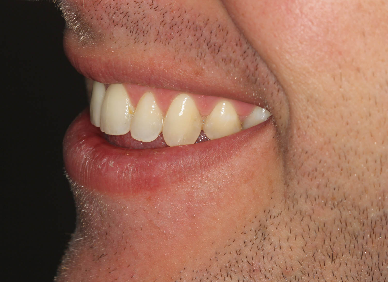
This is the result, after treatment. Can you pick the implant tooth?
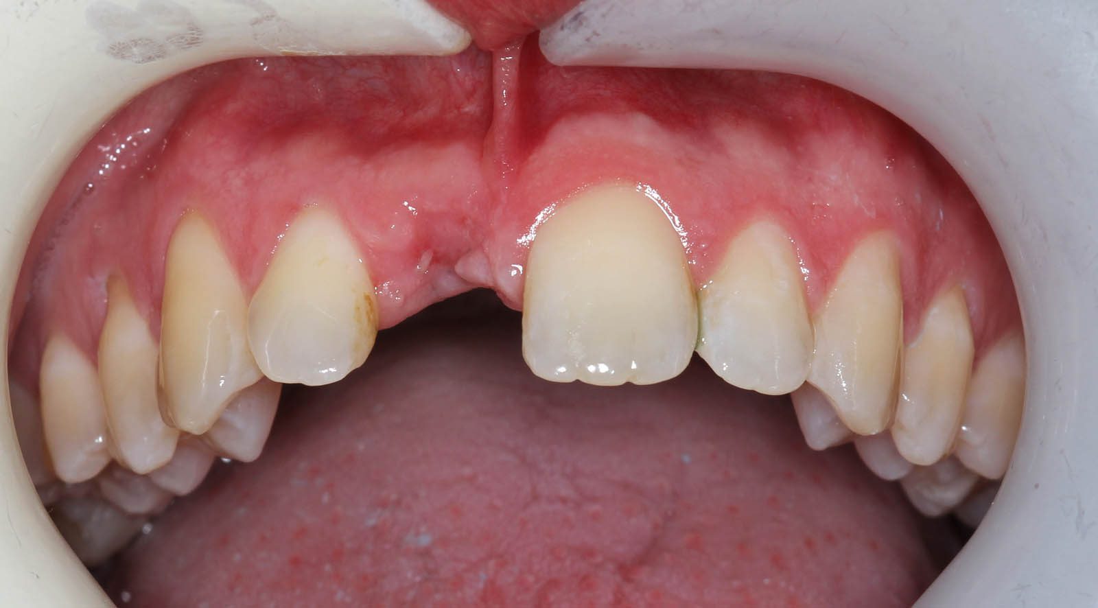
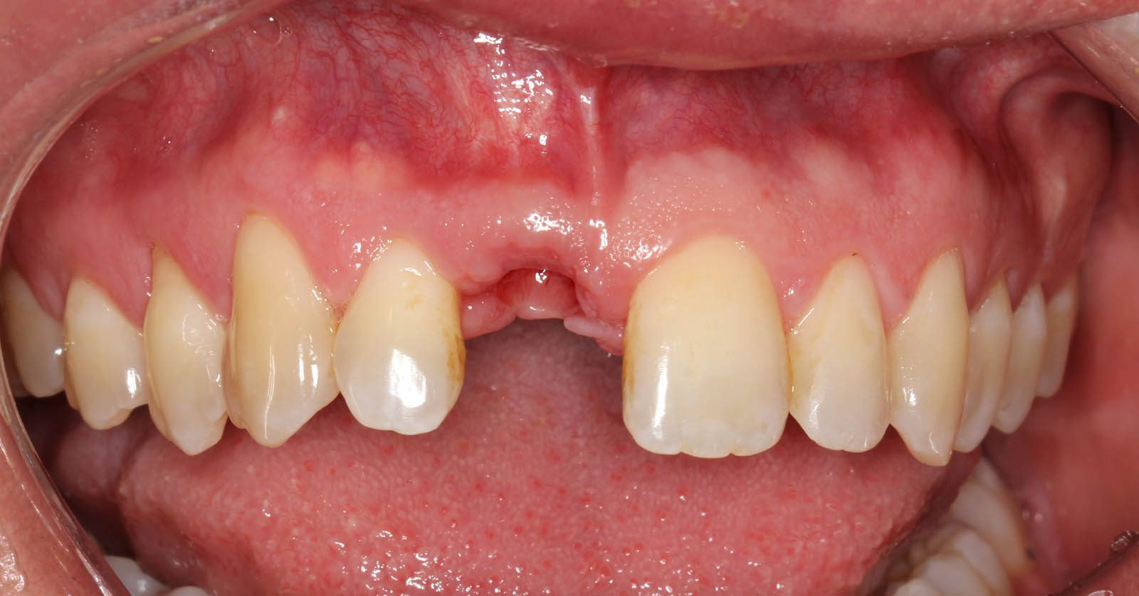
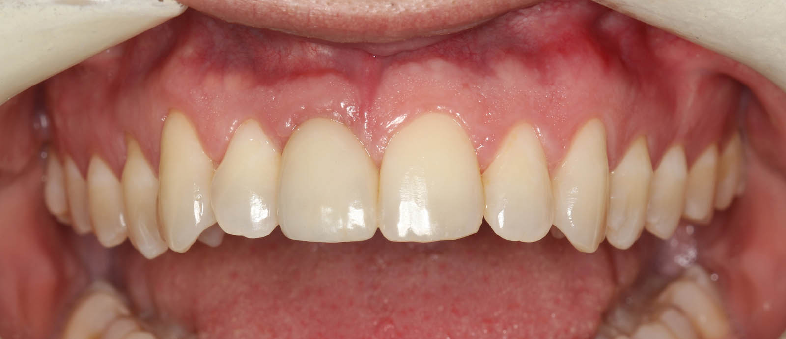
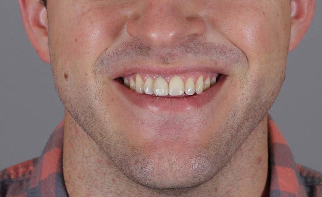
Pre-treatment frontal view. Missing #8 (upper right central incisor).
Pre-treatment, occlusal view. Note the concave nature (bony deficiency) compared to the adjacent teeth.
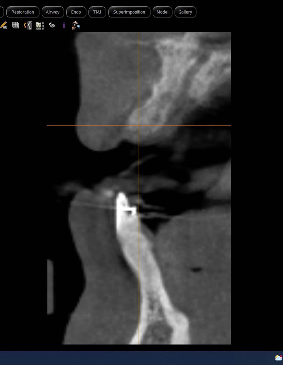
This x-ray is called a CBCT scan and was taken before the bone graft. When you compare it to the next CBCT scan you will see the dramatic bone growth that has happened since the bone graft surgery.
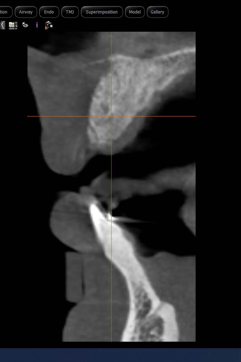
CBCT scan after 5 months of healing since the bone graft surgery. The amount of bone has doubled.
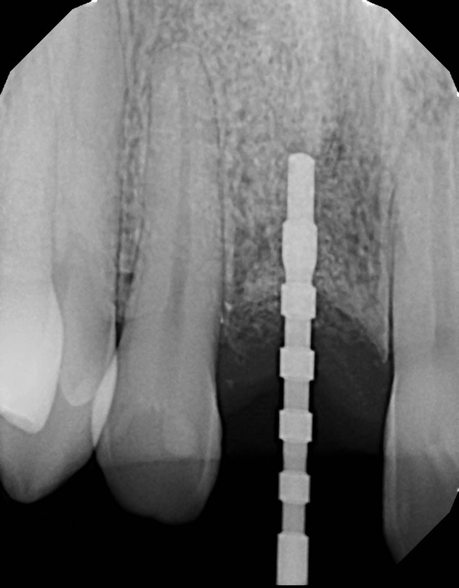
Guide pin x-ray taken at the time of implant placement surgery.
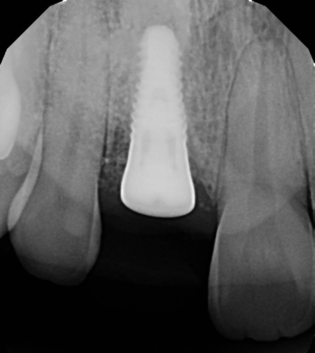
Implant in place, x-ray.
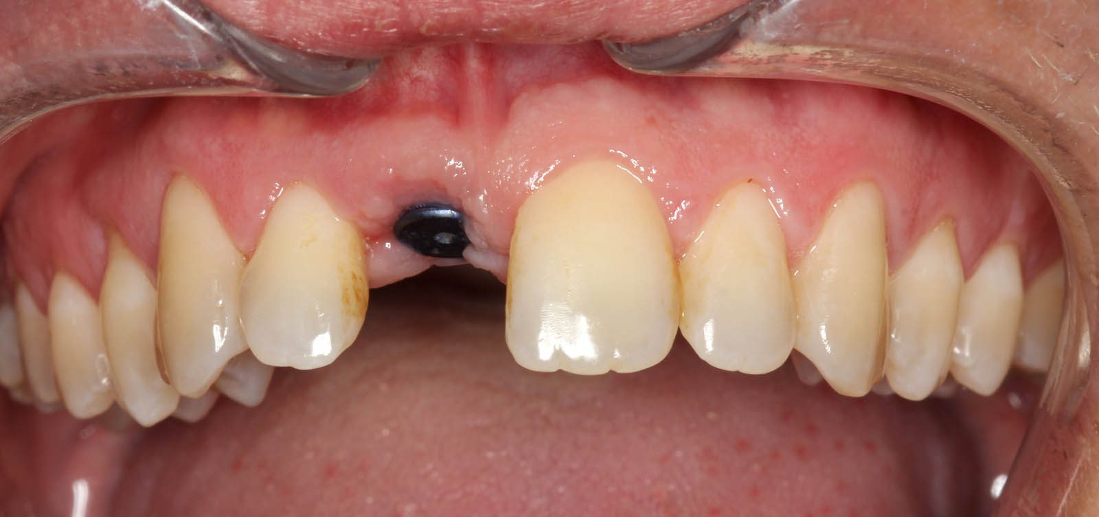
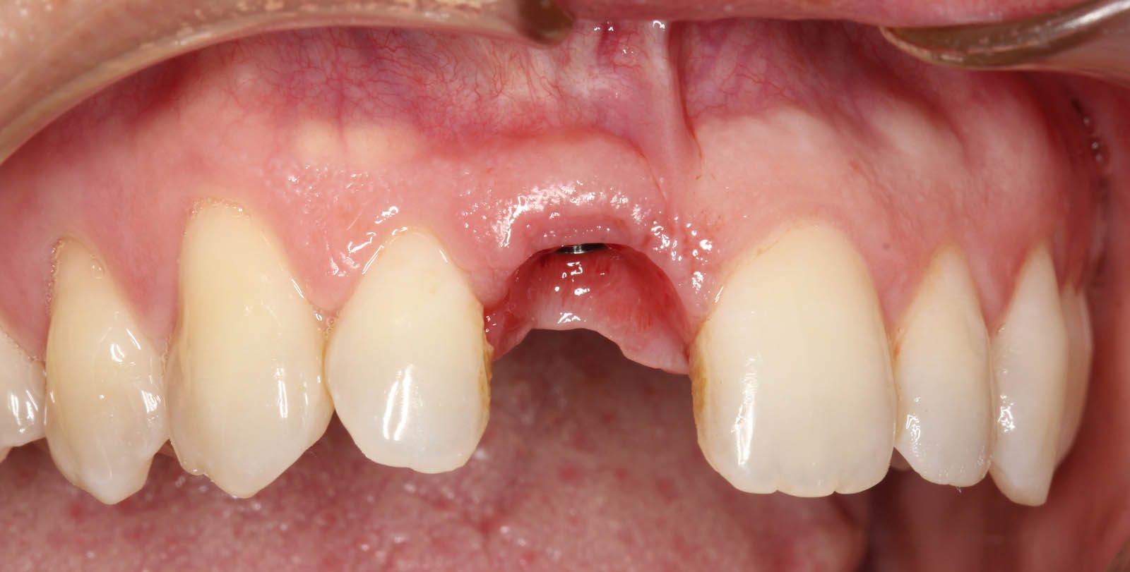
Two months after implant placement, healing abutment in place. The patient is ready for final impression of the implant.
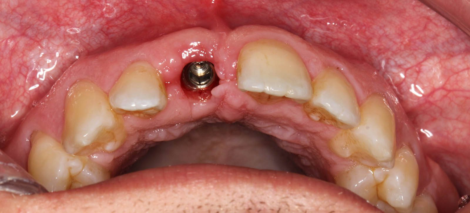
Two months after placement, without healing abutment, frontal view.
Two months after placement, without healing abutment, occlusal view.
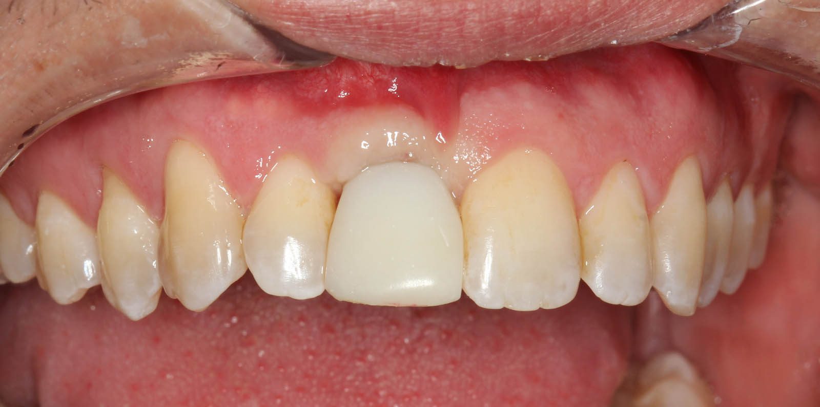
Temporary crown in place. Patient had in-office whitening done this day hence the mismatch of color.
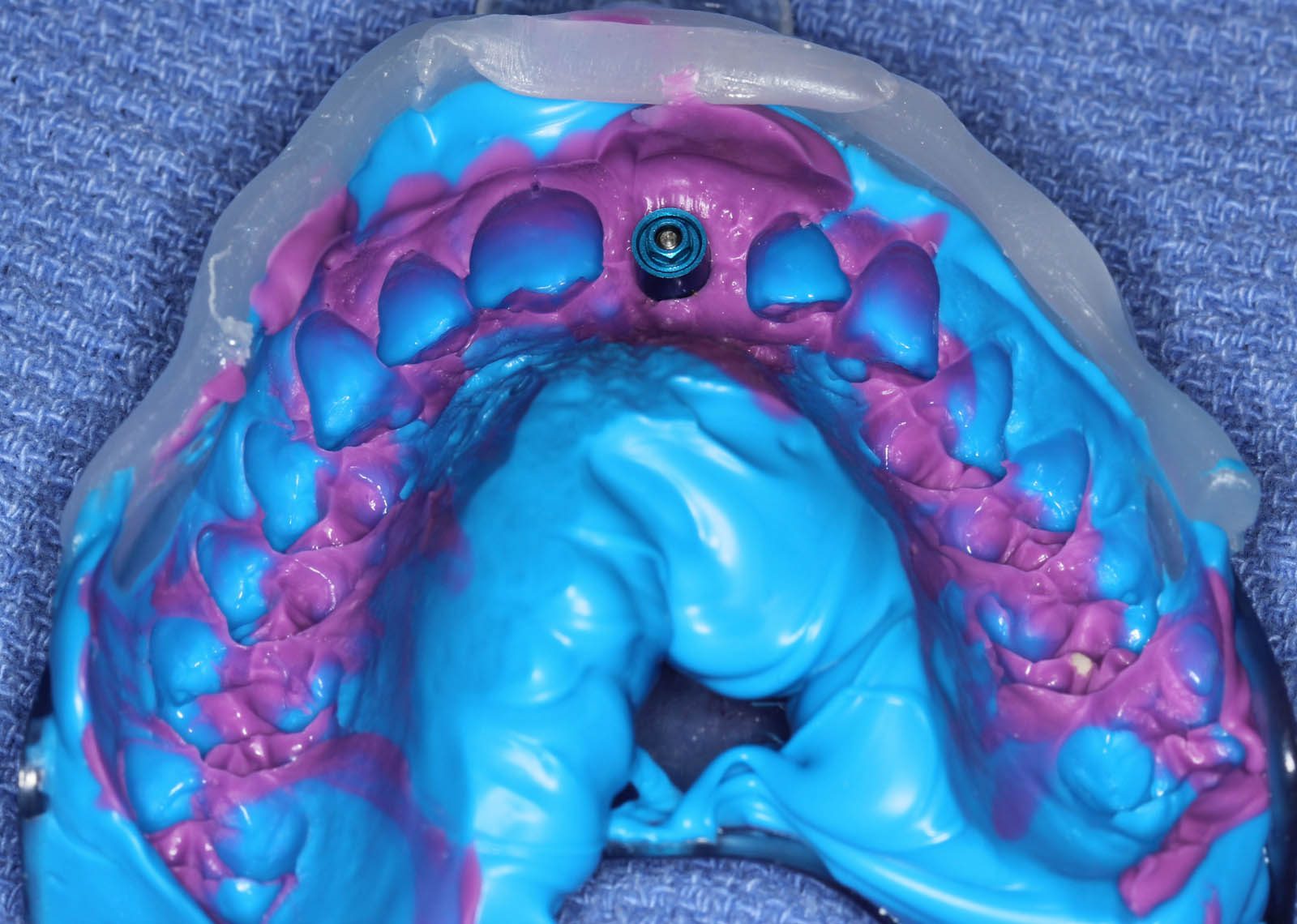
Final impression of the upper arch and implant.
Final impression with lab analog (replica of the implant) in place.
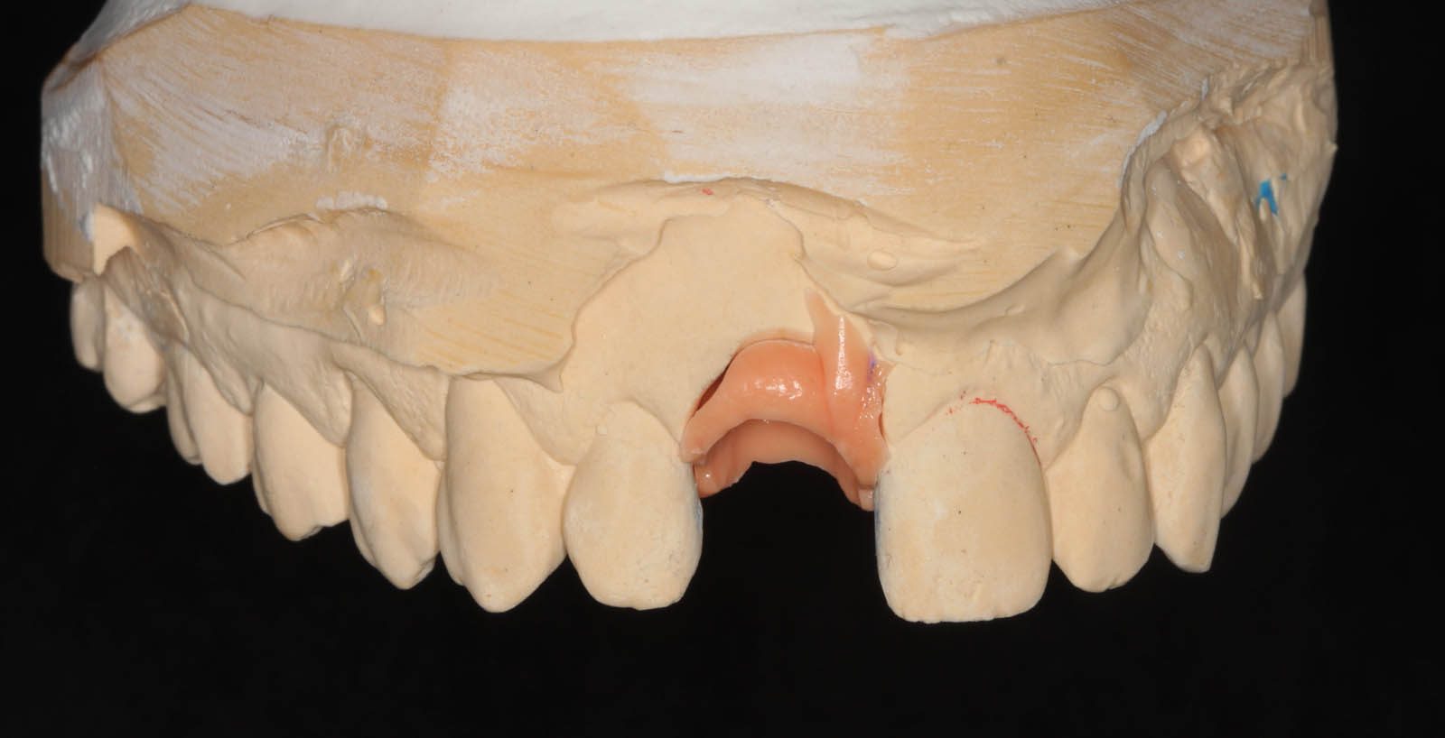
This is the “Master Cast” with soft tissue moulage (frontal view) made by pouring stone into the final impression.
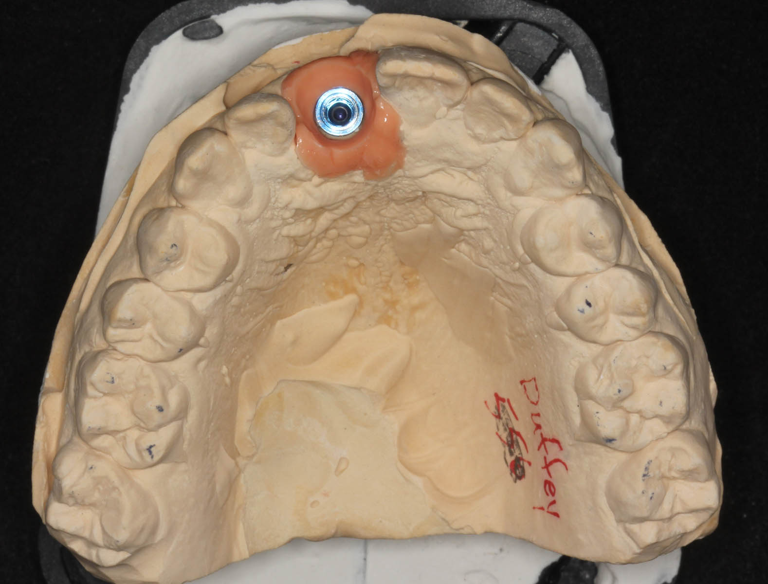
Master cast, occlusal view.
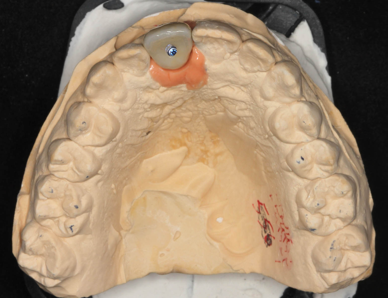
Master cast with screw-retained implant crown in place (occlusal view).
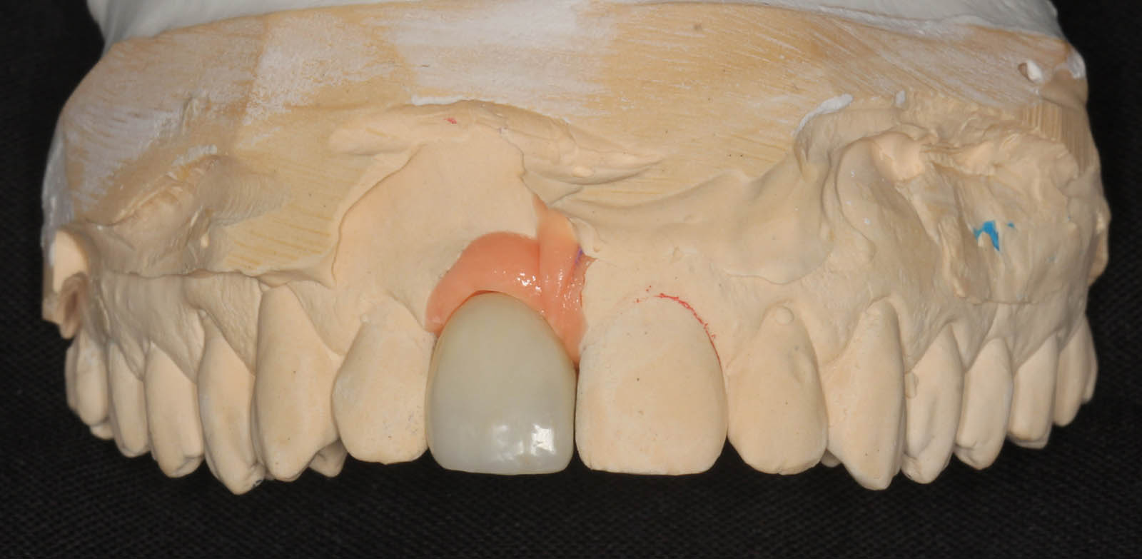
Master cast with screw-retained implant crown in place (frontal view).
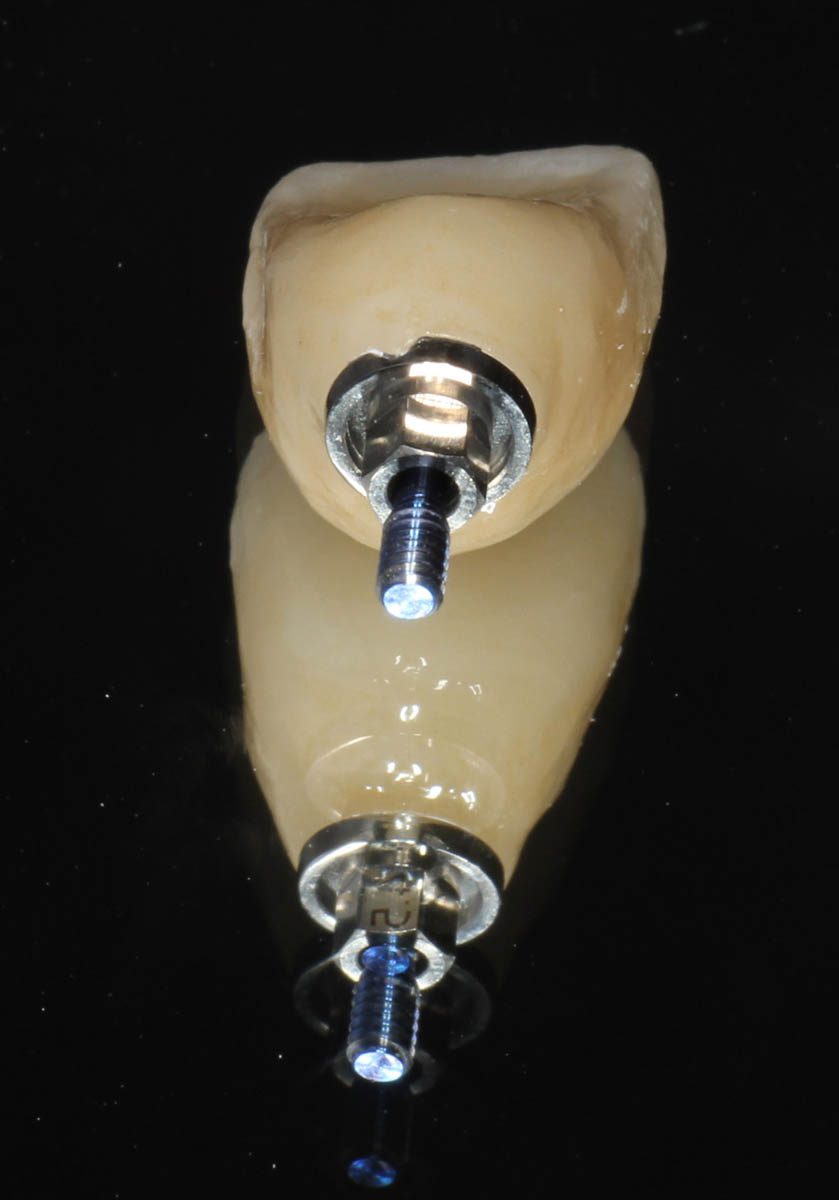
Screw-retained implant crown (tissue view). Note the interface that matches the implant analog which matches the implant.
Implant site with soft tissue development from the shape of the temporary crown. As you can expect, this is a perfect recipient site for the final crown.
Definitive (final) implant crown (frontal view).
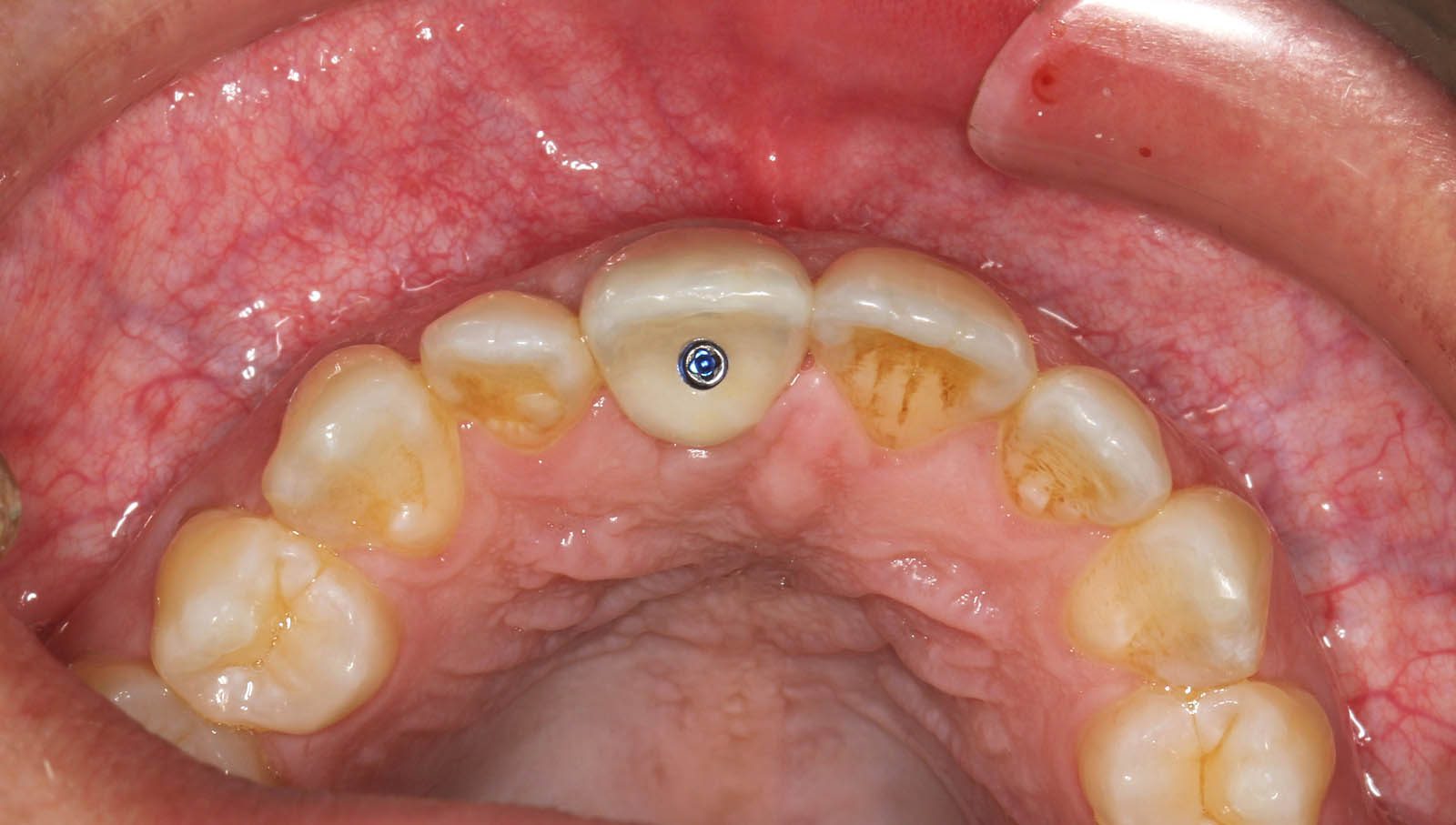
Definitive crown, occlusal view showing the retaining screw (blue).


 (Female)")