1 of 27
A screen shot of the panoramic view from a CT scan. The patient has almost no bone in the upper jaw. In order to have upper implants, he will need sinus and onlay bone grafting to increase bone height and width (total bone volume).
Upper jaw viewed from below. The patient lost his teeth some years early. He has been without teeth for several years. Bone volume is poor.
Lower jaw viewed from front. The patient lost his teeth some years early. He has been without teeth for several years. Bone volume is moderate to good.
Pre-treatment, portrait picture with reluctant smile. Note the reduced height of the lower 1/3rd of his face. Without teeth the jaw over-closes. In dentistry, we refer to this as a loss of Vertical Dimension of Occlusion (VDO).
Panoramic x-ray after sinus and onlay bone grafting. Thought subtle, you can see the ping-pong ball size sphere of new bone in the sinuses.
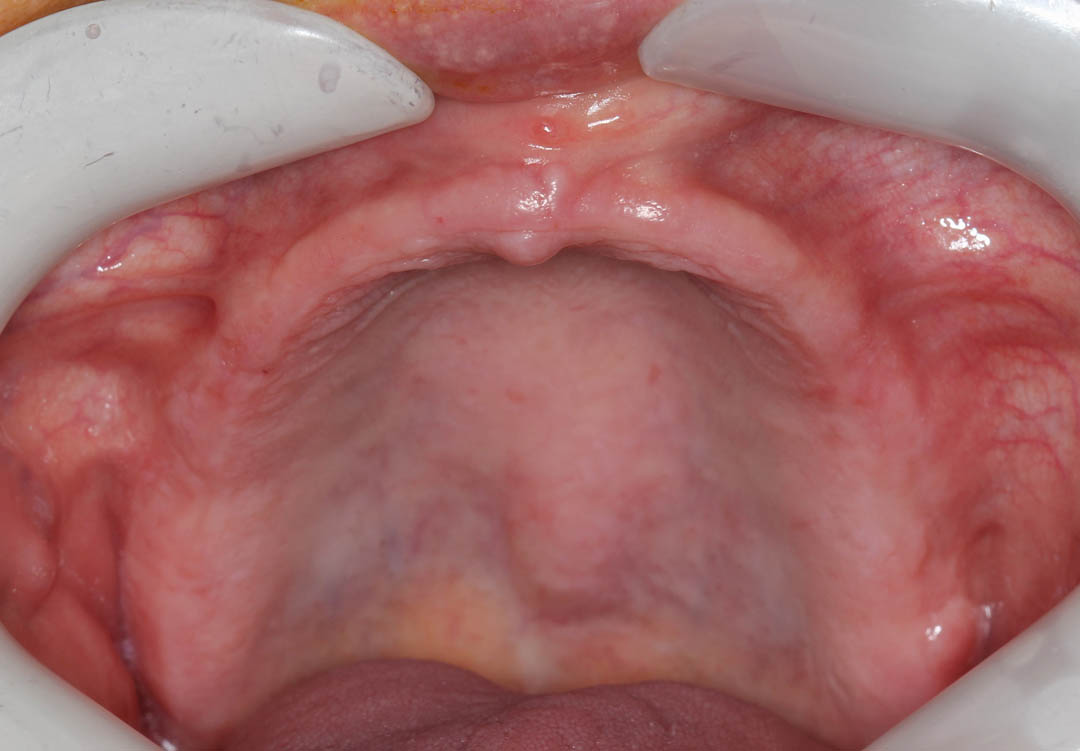
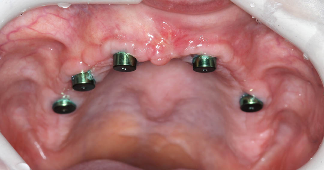
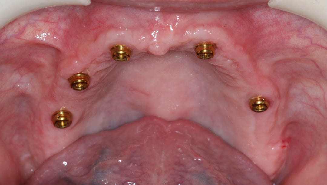
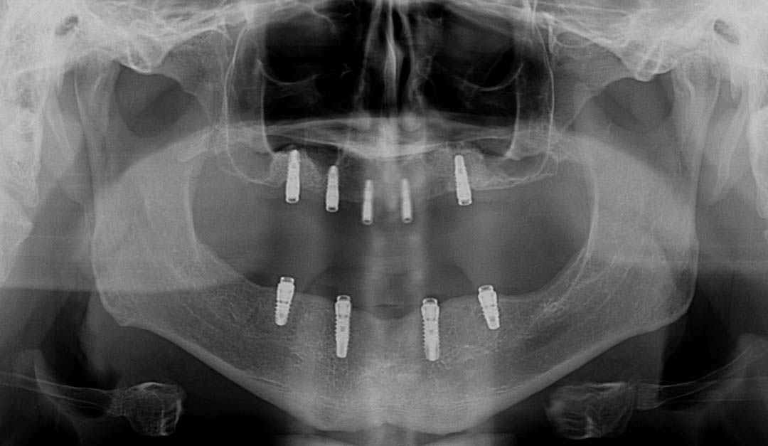
Upper jaw with healing caps over implants. Two months after implants are placed, the process of fabricating the overdentures begin. This process is nearly identical to making complete dentures (without implants). When I lecture, I tell the audience of dentists that, “implants only partially rescue poorly made dentures”! Meaning, for an optimum outcome, it requires great dentures connected to well placed implants.
Actual implant overdentures, frontal view of both. These are “digitally” fabricated through scanning a mock denture and milling these dentures out of a puck or PMMA - (Polymethylmethacrylate). This technology gives much better strength. Another great bonus is that it is very simple to make the patient a second set or replace broken dentures quickly.
Occlusal view of upper overdenture. Note, no palate. Patients rave about the return of taste and eating enjoyment.
Upper overdenture with attachments in place. (Different patient0. The color of the attachment signifies the amount of retention. There are numerous colors and numerous amounts of retention available. We spend significant time trying to find that amount (titration) that is ideal for each patient.